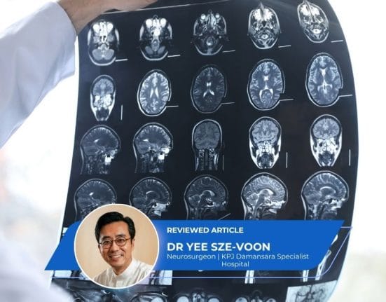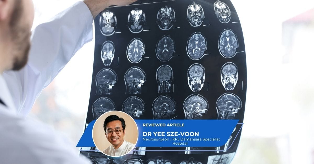
An incidental finding is an unexpected abnormality discovered on a brain Magnetic Resonance Imaging (MRI) or Computed Tomography (CT) scan. For example, a brain scan performed to investigate symptoms such as headache, dizziness, or a recent head injury may reveal an unexpected abnormality that requires further evaluation. The term incidentaloma is also used in clinical practice, generally referring to an incidentally discovered mass or lesion rather than every unexpected imaging finding.
Discovering an abnormal brain finding can naturally cause anxiety. However, many incidental brain findings are benign or have no immediate clinical consequence, although some require formal specialist assessment to establish their true significance.
Back To: Brain and Spine Care Main Page
Examples of Incidental Brain Findings
Incidental brain findings encompass a wide spectrum of structural, vascular and other abnormalities:
- White Matter Hyperintensities (WMHs): Areas of altered signal intensity on MRI scans that have several possible causes. In adults, WMHs can be markers of cerebrovascular and cardiovascular risk and may prompt assessment of factors such as hypertension, diabetes and other cardiovascular risk factors.
- Arachnoid Cysts: Fluid-filled sacs located between the brain tissue and the skull. Most are congenital and many remain asymptomatic, though their clinical behaviour can vary depending on location and size.
- Meningiomas: Tumours arising from the meninges (the protective membranes covering the brain and spinal cord). Most are benign and slow-growing, although some can behave more aggressively.
- Pituitary Incidentalomas: Lesions of the pituitary gland that are often asymptomatic but may require hormonal evaluation, visual assessment, or follow-up imaging depending on their size, location, and characteristics.
- Cerebral Aneurysms: Vascular abnormalities involving a bulge in a cerebral blood vessel wall. If detected incidentally, they may require specialist assessment to determine whether further imaging, monitoring, or treatment is appropriate.
- Developmental Venous Anomalies (DVAs): Variations in normal venous drainage. DVAs are generally benign and asymptomatic, usually requiring no active treatment, though associated vascular abnormalities or rare complications may require assessment.
What to Do After an Incidental Brain Finding Is Reported
If your radiology report indicates an unexpected brain finding, taking structured, deliberate steps ensures an appropriate clinical evaluation:
- Obtain Your Full Scan Package: Secure both the written radiological report and the digital image files (typically provided on a CD or USB flash drive in DICOM format) from the imaging provider.
- Consult Your Requesting Doctor: Review the report with the physician who initially ordered the scan to evaluate the results alongside your overall medical history.
- Seek Specialist Evaluation: Depending on the nature of the finding, schedule a consultation with an appropriate specialist—such as a neurologist or neurosurgeon. Depending on the finding, assessment may also involve endocrinology, ophthalmology, or a specialist in neurovascular conditions.
- Determine the Clinical Management Strategy: Depending on the specific diagnosis, management strategy falls into several potential pathways:
- Observation and Surveillance: If follow-up imaging is recommended, the timing depends strictly on the type, size, location, and characteristics of the finding, as well as the patient’s individual clinical circumstances. Some findings require interval imaging, while others may not require routine surveillance.
- Targeted Diagnostic Testing: Ordering specific laboratory investigations (such as hormone panels for pituitary lesions) or targeted vascular imaging (such as MR Angiography) as clinically indicated.
- Active Intervention: Recommending medical treatment, endovascular procedures, radiosurgery, or surgical intervention depending entirely on the diagnosis and clinical risk.
Navigating Specialist Consultations in Malaysia
When preparing to consult a medical specialist in Malaysia regarding an unexpected brain finding, organisation is key to an accurate clinical review.
What to Bring to Your Specialist Appointment:
- The radiologist’s written report
- The original CT or MRI images, preferably in DICOM format
- Previous brain imaging scans, if available for baseline comparison
- A list of current medications and medical history
- A summary of the symptoms or reasons that led to the original scan
Specialist Access and Hospital Accreditation: Depending on the private hospital or individual specialist’s referral policy, patients in Malaysia may be able to arrange a specialist consultation directly or through a primary care referral. When selecting a facility, patients may wish to check whether a hospital holds recognised healthcare accreditations, such as accreditation by the Malaysian Society for Quality in Health (MSQH). Accreditation involves assessment against established quality and safety standards and should be considered alongside the availability of relevant specialists and appropriate diagnostic infrastructure.
When to Seek Urgent Medical Attention
An incidental finding does not necessarily require emergency treatment. However, seek urgent medical attention if you develop symptoms such as:
- Sudden, severe headache, particularly if it reaches maximum intensity rapidly
- New weakness or numbness on one side of the body
- Difficulty speaking or understanding speech
- Sudden loss or significant change in vision
- A new seizure
- Loss of consciousness
- Sudden, severe dizziness accompanied by neurological symptoms
Neurosurgeons in Malaysia You May Consider
Choosing the right specialist is a critical step in your assessment and peace of mind. Some Neurosurgeons you may consider for comprehensive diagnosis and management:


Featured Doctor - Dr Yee Sze-Voon
For inquiries or for an introduction to a specialist, contact us below.
Frequently Asked Questions (FAQs)
1. Does an incidental brain finding mean I have brain cancer?
No. Many incidental findings discovered on brain MRI or CT scans are benign or have no immediate clinical consequence. Examples include some developmental cysts, benign tumours, and vascular variants. However, every finding requires individual interpretation by a doctor to determine if further monitoring or assessment is needed.
2. Should I immediately get a second MRI scan?
An immediate repeat scan is rarely necessary unless recommended by a specialist. Whether additional imaging is required depends on the quality of the original scan, the specific finding, and the recommendations of the radiologist or consulting specialist. If imaging surveillance is required, the timing will be tailored to the specific condition.
3. Can an abnormal MRI finding explain my headaches or dizziness?
It can, but not every abnormal finding is responsible for the symptoms that led to the scan. A specialist will consider the imaging findings alongside your symptoms, medical history, and neurological examination to determine whether there is a meaningful clinical connection.
4. What is the difference between seeing a neurologist and a neurosurgeon for an incidental lesion?
A neurologist focuses on the non-surgical evaluation, medical management, and long-term monitoring of neurological conditions. A neurosurgeon evaluates whether a structural lesion may require surgical or other procedural treatment. Some vascular conditions may also require assessment by a specialist trained in neurovascular or endovascular procedures.
5. How do specialists assess whether an incidental finding is changing over time?
Specialists evaluate sequential imaging by comparing the current and previous scans directly to assess whether the finding has remained stable or changed over time. Ensuring you bring your original imaging files (in DICOM format) to follow-up consultations allows your physician to make these direct visual comparisons.
For inquiries or for an introduction to a specialist, contact us below.

This medical content has been reviewed by Dr Yee Sze-Voon, a Consultant Neurosurgeon specialising in brain and spine surgery, minimally invasive procedures, and pain management. His commitment to precise diagnosis and patient-centred care makes him a trusted specialist for comprehensive neurological health.
All content on this site is informative only and is not intended to be a substitute for professional medical advice,
diagnosis, or treatment, and should never be relied upon for specific medical advice.
If you are experiencing a medical emergency, call 999 immediately or go to the nearest Accident & Emergency department.
365Asia is a member of Group 365Asia.